Contents
Scroll to:
https://doi.org/10.37489/2949-1924-0136
EDN: VJZOQW
Scroll to:
Background. 5-fluorouracil (5-FU) is a basic chemotherapeutic agent, but its clinical use is limited by rapid metabolism and high toxicity. The development of controlled release systems based on polymethacrylic acid (PMAA) allows for an improved pharmacokinetic profile of the drug, which requires validation of analytical methods for the resulting compound to ensure the quality requirements of such polymer systems.
Objective. Development and validation of a method for the quantitative determination of 5-FU in a PMAA based polymer complex using UV spectroscopy in various media simulating biological conditions.
Materials and methods. The object of the study was a lyophilized polymer complex of 5-FU and PMAA, obtained by liquid phase synthesis. The analysis was performed by UV spectroscopy on a Shimadzu UV-1800 instrument at a wavelength of 265–267 nm. The method was validated in accordance with the State Pharmacopoeia XV for the following parameters: specificity, linearity, accuracy, repeatability, and intralaboratory precision (with the participation of two operators). The studies were conducted in three media: purified water, phosphate buffer (pH 7.4 blood) and citrate buffer (pH 5.1 intracellular medium).
Results. Complex formation via hydrogen bonds between 5-FU and the carboxyl groups of PMAA was confirmed. The method was deemed specific, as the polymer carrier does not absorb in the analytical range (265 nm). A linear relationship was established (R[2] > 0.999). The recovery rate (accuracy) ranged from 98.48 % for a citrate buffer solution to 99.23 % for a phosphate buffer solution. The coefficient of variation (RSD) during precision testing did not exceed 2 %, confirming that the proposed method has satisfactory repeatability (convergence) and ensures reproducible results with multiple sample analyses under identical conditions. Calculated Student's and Fisher's t-tests confirmed the absence of systematic errors between operators. The study revealed that in an acidic environment (pH 5.1), the release of 5-FU was 15.92 % higher than at pH 7.4, confirming the pH sensitivity of the system.
Conclusions. The developed spectrophotometric method fully meets validity requirements and can be used for quality control of PMAA based polymeric 5-FU delivery systems. The demonstrated selectivity of 5-FU release in an acidic environment confirms the potential of using polymeric complexes for targeted delivery to tumor tissue.
Zhukova O.V., Dubovskaya N.A., Khokhlov A.L. Development and validation of a method for the determination of 5‑fluorouracil in a polymer complex based on polymethacrylic acid using UV spectroscopy. Patient-Oriented Medicine and Pharmacy. 2026;4(2):5-15. (In Russ.) https://doi.org/10.37489/2949-1924-0136. EDN: VJZOQW
5-Fluorouracil (5-FU) is one of the chemotherapeutic compounds widely used in the treatment of oncological diseases. 5-FU is extensively employed in clinical practice for the treatment of various solid tumors [1]. 5-FU is rapidly metabolized in the body; therefore, maintaining a high serum concentration of this drug is critically important for improving its therapeutic activity. However, the use of 5-FU at high concentrations is associated with a significant risk of severe toxic effects [2–4].
The literature presents numerous studies aimed at developing controlled-release systems for 5-FU that improve treatment outcomes [5–9].
Polymethacrylates are a widely available class of synthetic polymers. Polymethacrylic and polyacrylic acids are water-soluble polymers containing functional carboxyl groups that enable their modification.
A study published in 2011 proposed a universal model of pH-sensitive nanoparticles for selective drug delivery, with polymethacrylic acid (PMAA) as one of the key components [10]. In 2016, an investigation was conducted on the in vitro and in vivo antitumor properties of PMAA combined with gold-containing nanoparticles and doxorubicin via an acid-labile cysteine bond [11].
Previously, a polymer complex of 5-FU based on PMAA was developed, demonstrating alterations in the pharmacokinetics and biodistribution profile of 5-FU following its incorporation into the polymer complex [12].
Validation of analytical methods for the determination of drugs within polymeric particles (micelles, polymersomes) and complexes is of particular significance in modern pharmaceuticals; however, methodological aspects of validation for such systems are fragmentarily addressed in the scientific literature [13, 14]. Polymer complexes are actively being developed to improve bioavailability, stability, and controlled drug release, particularly in oncology and the therapy of neurodegenerative diseases. However, drug integration into the polymer matrix leads to analytical challenges: matrix effects, low concentrations of the active substance (often <1 μg/mL), and heterogeneity of drug distribution necessitate rigorous method validation to ensure accuracy, reproducibility, and selectivity.
The objective of this study is to develop and validate a method for the quantitative determination of 5-FU using ultraviolet spectroscopy (UV spectroscopy) in a PMAA-based polymer complex.
The study objects were samples of the experimental polymer complex of 5-FU, presented as a lyophilizate for solution preparation.
The polymer complex of 5-FU based on PMAA was obtained by liquid-phase synthesis according to a previously described method [15].
UV spectroscopy was employed as the method for quantitative determination of 5-FU content in the polymer complex.
Determination was conducted in accordance with the General Pharmacopoeia Monograph 1.2.1.1.0003 "Spectrophotometry in the Ultraviolet and Visible Regions" of the State Pharmacopoeia XV edition [16].
Since the polymer derivative of 5-FU represents 5-FU bound by hydrogen bonds to the carboxyl groups of PMAA, solutions of the initial substances were prepared for analysis.
0.00083% 5-FU solution. An accurately weighed quantity of 5-FU substance (~0.01 g) was quantitatively transferred to a 100 mL volumetric flask, dissolved in water, the flask volume was adjusted to the mark with the same solvent, and mixed. An 8.3 mL aliquot was taken, placed in a 100 mL volumetric flask, the volume was adjusted to the mark with solvent, and mixed.
0.01% PMAA solution. An accurately weighed quantity of PMAA (~0.01 g) was quantitatively transferred to a 100 mL volumetric flask, dissolved in water, the flask volume was adjusted to the mark with the same solvent, and mixed.
0.01% 5-FU polymer complex solution. An accurately weighed quantity of 5-FU polymer complex (~0.01 g) was quantitatively transferred to a 100 mL volumetric flask, dissolved in water, the flask volume was adjusted to the mark with the same solvent, and mixed.
Ultraviolet absorption spectra were obtained using a UV-1800 spectrophotometer (Shimadzu, Japan) in the wavelength range of 190–400 nm (date of last verification: 29.09.2025). The optical density of the obtained solutions was measured at a wavelength of 265 nm (for 5-FU determination) in a 1 cm cuvette.
The linear relationship between absorbance and sample concentration enables quantitative determination. Therefore, 5-FU content was also calculated spectrophotometrically, based on the Beer-Lambert-Bouguer law. The calibration curve was established by measuring the absorbance of several standard solutions of known concentration.
0.01% standard 5-FU solution in aqueous medium. An accurately weighed quantity of 5-FU substance (~0.01 g) was quantitatively transferred to a 100 mL volumetric flask, dissolved in water, the flask volume was adjusted to the mark with the same solvent, and mixed.
0.01% standard 5-FU solution in phosphate buffer (pH 7.4). An accurately weighed quantity of 5-FU substance (~0.01 g) was quantitatively transferred to a 100 mL volumetric flask, dissolved in phosphate buffer, the flask volume was adjusted to the mark with the same solvent, and mixed.
0.01% standard 5-FU solution in citrate buffer (pH 5.1). An accurately weighed quantity of 5-FU substance (~0.01 g) was quantitatively transferred to a 100 mL volumetric flask, dissolved in phosphate-citrate buffer, the flask volume was adjusted to the mark with the same solvent, and mixed.
From each of the above standard solutions, a 5 mL aliquot was taken and diluted 2-fold. Subsequently, 5 mL aliquots were sequentially taken from the obtained solutions and also diluted 2-fold. The procedure was repeated 5 times in total. To obtain intermediate concentrations, 3 mL aliquots from the second, third, fourth, and fifth dilutions were mixed with 1.5 mL of the corresponding solvent.
0.008% 5-FU polymer complex solution. An accurately weighed quantity of 5-FU polymer complex (~0.01 g) was quantitatively transferred to a 100 mL volumetric flask, dissolved in water, the flask volume was adjusted to the mark with the same solvent, and mixed. An 80 mL aliquot was taken, placed in a 100 mL volumetric flask, the volume was adjusted to the mark with solvent, and mixed.
0.01% 5-FU polymer complex solution. An accurately weighed quantity of 5-FU polymer complex (~0.01 g) was quantitatively transferred to a 100 mL volumetric flask, dissolved in water, the flask volume was adjusted to the mark with the same solvent, and mixed.
0.012% 5-FU polymer complex solution. An accurately weighed quantity of 5-FU polymer complex (~0.012 g) was quantitatively transferred to a 100 mL volumetric flask, dissolved in water, the flask volume was adjusted to the mark with the same solvent, and mixed.
Each solution was prepared three times in the corresponding solvent (water, phosphate buffer (pH 7.4), citrate buffer (pH 5.1)).
An accurately weighed quantity of 5-FU polymer complex (~0.01 g) was quantitatively transferred to a 100 mL volumetric flask, dissolved in the solvent, the flask volume was adjusted to the mark with the same solvent, and mixed.
The procedure was repeated 6 times.
Test solutions for the "Precision" parameter assessment were prepared according to the method used for accuracy determination. The procedure was performed independently by two researchers for three concentrations of 5-FU: 0.0056, 0.0070, and 0.0084 mg/mL. For each concentration, each researcher prepared six parallel samples.
The optical density of the prepared standard 5-FU solutions, as well as the test solutions of the 5-FU polymer complex for validation parameter assessment (accuracy, repeatability, precision) in different media, was measured at wavelengths from 265–267 nm (n=5), depending on the pH of the medium and the buffer solution used.
The 5-FU content was calculated using the calibration curve equations (formula (1)):
X = A/b, where (1)
X — 5-FU content, mg/mL;
A — optical density, arbitrary units;
b — linear regression coefficient of the corresponding calibration curve.
The proposed method for the quantitative determination of 5-FU in the polymer derivative was validated in accordance with the General Pharmacopoeia Monograph "Validation of Analytical Methods" (State Pharmacopoeia XV, GPM 1.1.0012) for the parameters of specificity, linearity, accuracy, and precision (repeatability (convergence), intermediate (intralaboratory) precision).
As a result of intermolecular interaction between 5-FU and the carboxyl groups of PMAA residues, a water-soluble polymer complex is formed (Fig. 1).

Figure 1. Intermolecular interaction of 5-FU with the carboxyl groups of PMAA
Note: R is the PMAA residue
The intermolecular interaction is due to the formation of hydrogen bonds between the electronegative fluorine atom in 5-FU and the hydrogen atom in the carboxyl residue of methacrylic acid, which has a partial positive charge and is covalently bonded to the electronegative oxygen atom. A hydrogen bond is also formed between the hydrogen atom in the –NH– group of the pyrimidine ring and the oxygen atom in the methacrylic acid residue, as well as between the oxygen atom and the hydrogen atom in the methacrylic acid residue. This mechanism indicates complete complementarity of 5-FU to the methacrylic acid residue. Each 5-FU molecule fully binds two methacrylic acid residues and partially binds a third residue.
UV spectroscopy data (Fig. 2) serve as a method confirming the formation of the intermolecular complex of 5-FU with the PMAA polymer.
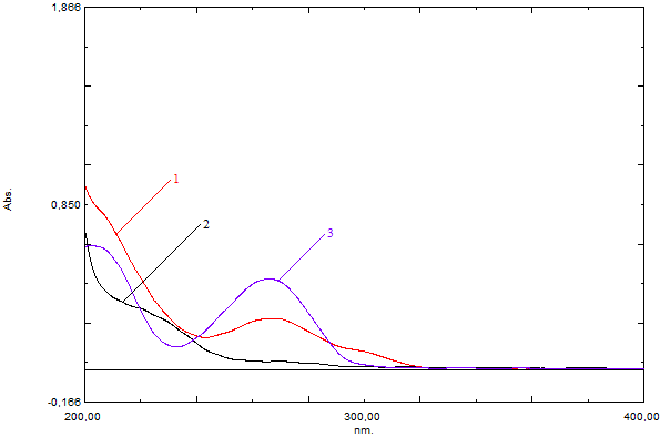
Figure 2. UV spectra of the 5-FU polymer complex (0.1 mg/mL), PMAA (0.1 mg/mL), and 5-FU (0.0083 mg/mL)
Notes: 1 — 5-FU polymer complex solution; 2 — PMAA solution; 3 — solution containing 5-FU substance.
To evaluate the potential of using the 5-FU polymer complex as a delivery system, an analysis of 5-FU release in media simulating biological environments was conducted.
A distinctive feature of interpolymer complexes and conjugates of antitumor drugs is their stability in a medium simulating the bloodstream environment (pH 7.35–7.45) and the decomposability of the complex/conjugate with the release of the antitumor drug in media simulating the intracellular space (pH 6.8–7.2), the tumor microenvironment (pH 6.2–6.8), or the pH of intracellular lysosomes (pH 4.0–5.0).
Two buffer solutions were used as media simulating biological environments: phosphate buffer (PB) at pH 7.4 — to simulate the bloodstream, and citrate buffer (CB) at pH 5.1 — to simulate the intracellular space.
It was found that the release of the polymer complex with 5-FU in an acidic medium (at pH 5.1) was 15.92% higher than at pH 7.4. This observation is explained by accelerated hydrolysis of pH-sensitive bonds in the polymer matrix at reduced pH values; changes in electrostatic interactions between the carrier and 5-FU; and increased polymer swelling under acidic conditions, which facilitates 5-FU diffusion.
The obtained data have important practical significance for the development of targeted delivery systems for antitumor drugs.
Therefore, the study involved validation of the quantitative determination method for 5-FU by UV spectroscopy in different media: water; phosphate buffer (pH 7.4); and citrate buffer (pH 5.1).
Method specificity was determined by comparing the UV absorption spectra of the test samples and possible interfering components in the solvents used.
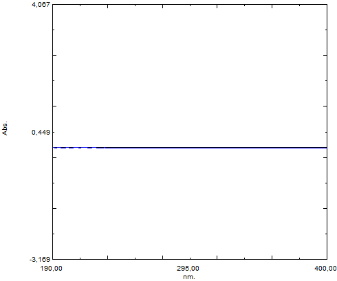
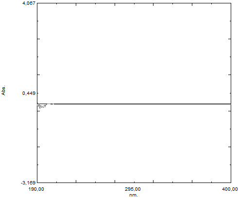
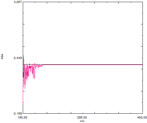
Initially, UV spectra of all solvents used in the method (purified water, phosphate (pH 7.4) and citrate (pH 5.1) buffers) were recorded in the 190–400 nm range (Fig. 3).
 |  |  |
| purified water | phosphate (pH 7.4) buffers | citrate (pH 5.1) buffers |
Figure 3. UV spectra of the solvents used
It was established that none of the solvents absorb light in the 260–265 nm region, meaning their own contribution to the signal is absent.
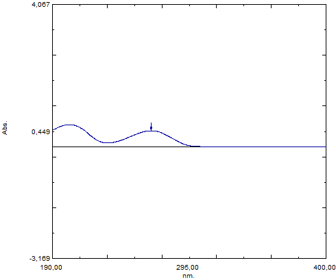
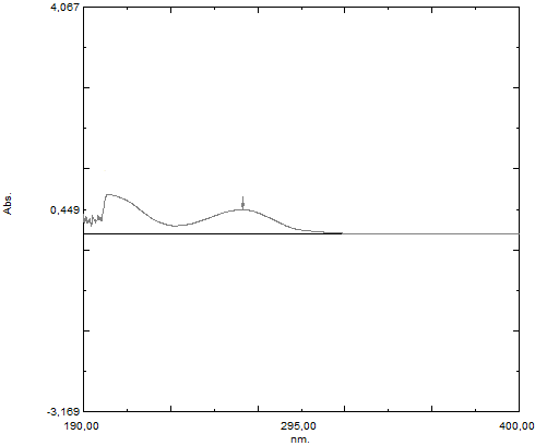
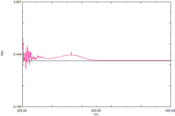
UV spectra of standard 5-FU solutions in all media used showed a clear absorption maximum at 265–267 nm (Fig. 4). This confirmed that this absorption band is characteristic of 5-FU and can be used as an analytical signal.
 |  |  |
| purified water | phosphate (pH 7.4) buffers | citrate (pH 5.1) buffers |
Figure 4. UV spectra of 5-FU solutions (0.0083 mg/mL)
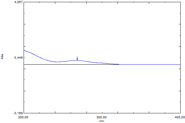
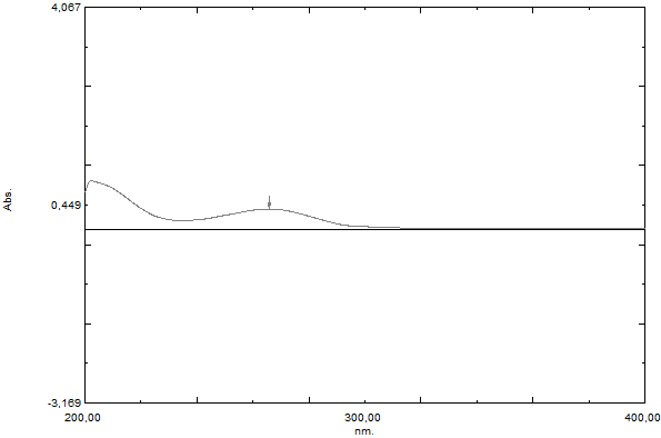
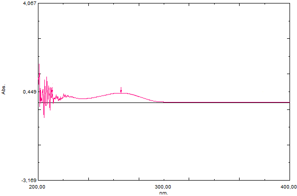
To confirm that the absorption band at 265–267 nm in the solution of the test 5-FU polymer complex belongs specifically to the drug molecule rather than the polymer or impurities, its UV spectra were recorded (Fig. 5).
 |  |  |
| purified water | phosphate (pH 7.4) buffers | citrate (pH 5.1) buffers |
Figure 5. UV spectra of 5-FU polymer complex solutions (0.1 mg/mL)
The absorption spectra of the 5-FU polymer complex coincided with those of pure 5-FU, showing the same maximum at 265–267 nm depending on the medium. The coincidence of the spectra proves that the proposed spectrophotometric method is specific for the determination of 5-FU in the polymer complex. The polymer carrier does not interfere with the measurement of 5-FU concentration in the selected wavelength range.
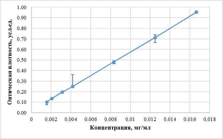
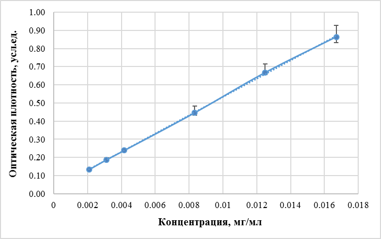
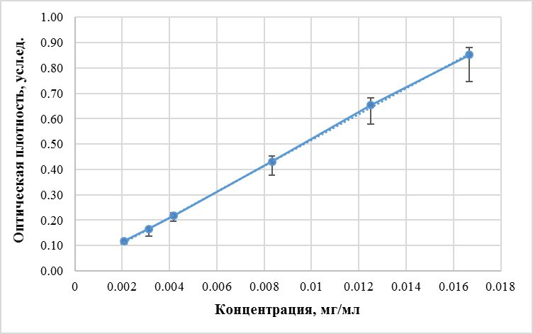
Method linearity was evaluated in the working concentration range of 5-FU from 0.0016 to 0.0167 mg/mL.
Prior to statistical processing of data for constructing the calibration dependence, homogeneity of measurements was tested for each prepared concentration of standard solutions. The Q-test was used to detect gross errors in small samples. At a confidence probability of p=0.99, all calculated Q-test values for the experimental data did not exceed the critical table value Qcrit. (0.99; n=0.760). This confirmed sample homogeneity and the absence of statistical outliers, allowing further analysis.
The dependence of optical density on 5-FU concentration in the indicated range is well described by a linear equation of the form y = bx + a (Fig. 6).
 |  |  |
| purified water | phosphate (pH 7.4) buffers | citrate (pH 5.1) buffers |
Figure 6. Calibration graphs for linearity study of the method for quantitative determination of 5-FU by UV spectroscopy
Significance of the coefficients was tested using Student's t-test (Table 1).
Table 1. Significance of regression coefficients a, b
| Medium | t(calc.)b | Standard error, S_b | t(calc.)a | Standard error, S_a | t(tab.) |
|---|---|---|---|---|---|
| Water | |||||
| 206.436 | 0.2713 | 6.8043 | 0.0024 | 4.03 | |
| Phosphate buffer (pH 7.4) | |||||
| 112.4741 | 0.4478 | 6.6811 | 0.0042 | 4.60 | |
| Citrate buffer (pH 5.1) | |||||
| 109.9668 | 0.4629 | 1.9277 | 0.0044 | 4.60 |
It was established that coefficients a and b are statistically significant for water and phosphate buffer (pH 7.4). The final linear equation for water is y=56.01x+0.0162; for phosphate buffer (pH 7.4) — y=50.365x+0.029. For citrate buffer (pH 5.1), coefficient *b* was found to be statistically significant, while coefficient a was not. Based on this, the calibration dependence was reconstructed as the equation y=bx passing through the origin. The final linear equation for citrate buffer is y=51.645x. The coefficient of determination R² is 0.9994.
Statistical processing of experimental data for the linearity study was performed (Table 2).
Table 2. Results of statistical processing of experimental data obtained in the linearity study for the dependence y = bx + a
| f | x̄ | ȳ | b | a | t(99,3) | Δb | Δa | S₀² | r |
|---|---|---|---|---|---|---|---|---|---|
| Water | |||||||||
| 5 | 0.0069 | 0.4037 | 56.01 | 0.0162 | 4.03 | 1.093 | 0.0163 | 0.000015 | 0.99995 |
| Phosphate buffer (pH 7.4) | |||||||||
| 4 | 0.0078 | 0.42 | 50.365 | 0.029 | 4.60 | 2.0598 | 0.0193 | 0.000034 | 0.9998 |
| Citrate buffer (pH 5.1) | |||||||||
| 4 | 0.0078 | 0.4061 | 51.645 | — | 4.60 | 2.1293 | — | 0.000069 | 0.9997 |
Method accuracy was evaluated from 9 determinations at 3 concentration levels (0.0056 mg/mL; 0.0070 mg/mL; 0.0084 mg/mL). The mean recovery of 5-FU based on quantitative determination results in water was 98.91%, in phosphate buffer (pH 7.4) was 99.23%, and for the method in citrate buffer (pH 5.1) was 98.48% (Table 3).
Table 3. Metrological characteristics of the quantitative determination method for 5-FU in the polymer complex using UV spectroscopy
| n | f | μ | x̄, % | S² | S | P, % | T(P,f) | ΔX | Δx̄ | ε̄, % | RSD, % |
|---|---|---|---|---|---|---|---|---|---|---|---|
| Water | |||||||||||
| 9 | 8 | 100 | 98.91 | 2.0617 | 1.4359 | 99 | 3.36 | 4.8245 | 1.6082 | 1.6259 | 1.4518 |
| Phosphate buffer (pH 7.4) | |||||||||||
| 9 | 8 | 100 | 99.23 | 2.0553 | 1.4336 | 99 | 3.36 | 4.8169 | 1.6057 | 1.6181 | 1.4447 |
| Citrate buffer (pH 5.1) | |||||||||||
| 9 | 8 | 100 | 98.48 | 0.5284 | 0.7269 | 99 | 3.36 | 2.4425 | 0.8142 | 0.8267 | 0.7381 |
The analytical range of the method was evaluated based on the results of "Linearity" and "Accuracy" assessments. Acceptance criterion: for quantitative determination of the main substance, the method range should be 80–120% of the nominal content.
Based on the results obtained for the "Linearity" and "Accuracy" parameters, the method range is 80–120%, as "Accuracy" was determined at concentrations of 80% (0.0800 mg/mL of 5-FU polymer complex and 0.0056 mg/mL of 5-FU), 100% (0.1000 mg/mL of 5-FU polymer complex and 0.007 mg/mL of 5-FU), and 120% (0.1200 mg/mL of 5-FU polymer complex and 0.0084 mg/mL of 5-FU) of the nominal content. "Linearity" was determined in the range of 22.33–238.57% of the standard sample concentration used in the analytical method.
Conclusion: the validated parameter meets the acceptance criterion.
Precision was investigated on homogeneous samples and evaluated in two variants: repeatability (convergence) and intralaboratory (intermediate) precision.
Repeatability (Convergence).
Repeatability was evaluated from six parallel determinations at a known 5-FU concentration (Table 4). All measurements were performed by a single operator on a single instrument over a short period of time.
Table 4. Metrological characteristics for determining repeatability of the quantitative determination of 5-FU by spectroscopy
| n | f | x̄, % | S | P, % | T(P,f) | ΔX | Δx̄ | RSD, % |
|---|---|---|---|---|---|---|---|---|
| Water | ||||||||
| 6 | 5 | 99.76 | 1.5189 | 99 | 4.03 | 6.1214 | 2.4991 | 1.5226 |
| Phosphate buffer (pH 7.4) | ||||||||
| 6 | 5 | 98.19 | 1.4509 | 99 | 4.03 | 5.8473 | 2.3871 | 1.4777 |
| Citrate buffer (pH 5.1) | ||||||||
| 6 | 5 | 97.46 | 1.0448 | 99 | 4.03 | 4.2106 | 1.7189 | 1.0720 |
The obtained RSD value did not exceed 2%, confirming that the proposed method exhibits satisfactory repeatability (convergence) and ensures reproducible results upon repeated sample analysis under identical conditions.
Intralaboratory Precision.
Intralaboratory precision was assessed to evaluate the reproducibility of method results under conditions allowing some variation within a single laboratory. The study was conducted at the Department of Pharmaceutical Chemistry and Pharmacognosy of Privolzhsky Research Medical University over a short period using the same analytical equipment.
The key varying factor in this study was the operator. Analysis was performed by two different researchers, each conducting a series of determinations for three different concentrations of standard 5-fluorouracil solution: 0.0056 mg/mL; 0.0070 mg/mL; 0.0084 mg/mL.
For each concentration and each operator, basic statistical parameters were calculated (Table 5).
Table 5. Metrological characteristics for determining intralaboratory precision in the quantitative determination of 5-FU by spectroscopy
| Concentration, mg/mL | Operator | μ | f | x̄, % | S | P | t(tab.) | Δx | ε | t(calc.) | F(99,5,5) | F(calc.) |
|---|---|---|---|---|---|---|---|---|---|---|---|---|
| Water | ||||||||||||
| 0.0056 | 1 | 100 | 5 | 98.19 | 1.5802 | 99 | 4.03 | 6.3683 | 6.4859 | 2.8117 | 3.45 | 1.0838 |
| 2 | 100 | 5 | 98.35 | 1.5179 | 99 | 4.03 | 6.1171 | 6.2200 | 2.6699 | 3.45 | ||
| 0.0070 | 1 | 100 | 5 | 99.76 | 1.5189 | 99 | 4.03 | 6.1214 | 6.1361 | 0.3853 | 3.45 | 0.8159 |
| 2 | 100 | 5 | 98.74 | 1.6816 | 99 | 4.03 | 6.7767 | 6.8631 | 1.8341 | 3.45 | ||
| 0.0084 | 1 | 100 | 5 | 98.89 | 1.3692 | 99 | 4.03 | 5.5180 | 5.5795 | 1.9711 | 3.45 | 0.7306 |
| 2 | 100 | 5 | 99.00 | 1.6019 | 99 | 4.03 | 6.4556 | 6.5205 | 1.5224 | 3.45 | ||
| Phosphate buffer (pH 7.4) | ||||||||||||
| 0.0056 | 1 | 100 | 5 | 98.27 | 1.9214 | 99 | 4.03 | 7.7431 | 7.8794 | 2.2046 | 3.45 | 1.2102 |
| 2 | 100 | 5 | 98.68 | 1.7466 | 99 | 4.03 | 7.0387 | 7.1326 | 1.8451 | 3.45 | ||
| 0.0070 | 1 | 100 | 5 | 99.09 | 1.3623 | 99 | 4.03 | 5.4899 | 5.5406 | 1.6431 | 3.45 | 1.1001 |
| 2 | 100 | 5 | 98.38 | 1.2988 | 99 | 4.03 | 5.2341 | 5.3205 | 3.0608 | 3.45 | ||
| 0.0084 | 1 | 100 | 5 | 99.59 | 1.0952 | 99 | 4.03 | 4.4135 | 4.4316 | 0.9160 | 3.45 | 1.4505 |
| 2 | 100 | 5 | 99.28 | 0.9093 | 99 | 4.03 | 3.6646 | 3.6914 | 1.9522 | 3.45 | ||
| Citrate buffer (pH 5.1) | ||||||||||||
| 0.0056 | 1 | 100 | 5 | 97.85 | 1.7899 | 99 | 4.03 | 7.2137 | 7.3720 | 2.9393 | 3.45 | 1.0565 |
| 2 | 100 | 5 | 100.91 | 1.7415 | 99 | 4.03 | 7.0181 | 6.9551 | 1.2749 | 3.45 | ||
| 0.0070 | 1 | 100 | 5 | 98.38 | 1.7111 | 99 | 4.03 | 6.8959 | 7.0092 | 2.3158 | 3.45 | 0.9341 |
| 2 | 100 | 5 | 99.90 | 1.7705 | 99 | 4.03 | 7.1350 | 7.1419 | 0.1333 | 3.45 | ||
| 0.0084 | 1 | 100 | 5 | 98.66 | 1.1843 | 99 | 4.03 | 4.7731 | 4.8379 | 2.7736 | 3.45 | 0.5126 |
| 2 | 100 | 5 | 97.39 | 1.6542 | 99 | 4.03 | 6.6665 | 6.8451 | 3.8631 | 3.45 |
Reproducibility was assessed using Student's t-test. Since t(calc.) < T(99%, 5), the results of the two samples are considered free from systematic error and therefore reproducible.
It has been demonstrated that the developed method satisfies validation requirements for the parameters of specificity, linearity, accuracy, and precision (repeatability (convergence), intermediate (intralaboratory) precision) in accordance with the General Pharmacopoeia Monograph "Validation of Analytical Methods" (State Pharmacopoeia XV, GPM 1.1.0012).
1. Diasio RB, Harris BE. Clinical pharmacology of 5-fluorouracil. Clin Pharmacokinet. 1989 Apr;16(4): 215-37. doi: 10.2165/00003088-198916040-00002
2. Kennedy BJ. 5-fluorouracil toxicity: old or new? Cancer. 1999 Oct 1;86(7):1099-100.
3. Alter P, Herzum M, Soufi M, et al. Cardiotoxicity of 5-fluorouracil. Cardiovasc Hematol Agents Med Chem. 2006 Jan;4(1):1-5. doi: 10.2174/187152506775268785.
4. Naren G, Guo J, Bai Q, et al. Reproductive and developmental toxicities of 5-fluorouracil in model organisms and humans. Expert Rev Mol Med. 2022 Jan 31;24:e9. doi: 10.1017/erm.2022.3.
5. Szota M, Reczyńska-Kolman K, Pamuła E, et al. Poly(amidoamine) Dendrimers as Nanocarriers for 5-Fluorouracil: Effectiveness of Complex Formation and Cytotoxicity Studies. Int J Mol Sci. 2021 Oct 16;22(20):11167. doi: 10.3390/ijms222011167.
6. Wang W, Joyce P, Bremmell K, et al. Liposomal 5-Fluorouracil Polymer Complexes Facilitate Tumor-Specific Delivery: Pharmaco-Distribution Kinetics Using Microdialysis. Pharmaceutics. 2022 Jan 18;14(2):221. doi: 10.3390/pharmaceutics14020221.
7. Mashaqbeh H, Obaidat R, Al-Shar'i NA, et al. Weak complexation of 5-fluorouracil with β-cyclodextrin, carbonate, and dianhydride crosslinked β-cyclodextrin: in vitro and in silico studies. Res Pharm Sci. 2022 Jul 14;17(4):334-349. doi: 10.4103/1735-5362.350235.
8. Szota M, Wolski P, Carucci C, et al. Effect of Ionization Degree of Poly(amidoamine) Dendrimer and 5-Fluorouracil on the Efficiency of Complex Formation-A Theoretical and Experimental Approach. Int J Mol Sci. 2023 Jan 3;24(1):819. doi: 10.3390/ijms24010819.
9. Grant JJ, Pillai SC, Perova TS, et al. Enhancement of 5-Fluorouracil Drug Delivery in a Graphene Oxide Containing Electrospun Chitosan/Polyvinylpyrrolidone Construct. Materials (Basel). 2024 Oct 31;17(21):5300. doi: 10.3390/ma17215300/
10. Tang H, Guo J, Sun Y, et al. Facile synthesis of pH sensitive polymer-coated mesoporous silica nanoparticles and their application in drug delivery. Int J Pharm 2011;421:388-96.
11. Yilmaz G, Demir B, Timur S, Becer CR. Poly (methacrylic acid)- coated gold nanoparticles: functional platforms for theranostic applications. Biomacromolecules 2016;17:2901-11.
12. Zhukova OV, Dubovskaya NA, Zykova DA, et al. Specifics of Pharmacokinetics and Biodistribution of 5-Fluorouracil Polymeric Complex. Molecules. 2023 Dec 15;28(24):8096. doi: 10.3390/molecules28248096.
13. Razavi L, Raissi H, Farzad F. Validation of an MD simulation approach for electrical field responsive micelles and their application in drug delivery. Sci Rep. 2023 Feb 15;13(1):2665. doi: 10.1038/s41598-023-29835-y.
14. Galbis E, Iglesias N, Lucas R, Tinajero-Díaz E, de-Paz MV, Muñoz-Guerra S, Galbis JA. Validation of Smart Nanoparticles as Controlled Drug Delivery Systems: Loading and pH-Dependent Release of Pilocarpine. ACS Omega. 2018 Jan 31;3(1):375-382. doi: 10.1021/acsomega.7b01421.
15. Dubovskaya N.A., Zhukova O.V. Preparation and evaluation of a complex of methacrylic acid polymer and 5-fluorouracil under different pH conditions. Modern problems of natural sciences and pharmacy. Collection of articles of the All-Russian scientific conference; May 20–23, 2025; Mari State University, Yoshkar-Ola; 2025; (14): 332–335.
16. State Pharmacopoeia of the Russian Federation. 15th edition. Moscow, 2023.
Olga V. Zhukova — Dr. Sci. (Pharm.), Associate Professor, Head of the Department of Pharmaceutical Chemistry and Pharmacognosy
Nizhniy Novgorod
The authors declare no conflict of interest
Natalya A. Dubovskaya — Assistant Professor, Department of Pharmaceutical Chemistry and Pharmacognosy
Nizhniy Novgorod
The authors declare no conflict of interest
Aleksandr L. Khokhlov — Dr. Sci. (Med.), Professor, Academician of the Russian Academy of Sciences, Head of the Department of Pharmacology and Clinical Pharmacology, Rector of the Yaroslavl State Medical University, Chairman of the Ethics Council under the Ministry of Health of the Russian Federation
Yaroslavl
The authors declare no conflict of interest
Zhukova O.V., Dubovskaya N.A., Khokhlov A.L. Development and validation of a method for the determination of 5‑fluorouracil in a polymer complex based on polymethacrylic acid using UV spectroscopy. Patient-Oriented Medicine and Pharmacy. 2026;4(2):5-15. (In Russ.) https://doi.org/10.37489/2949-1924-0136. EDN: VJZOQW
НАШИ КНИГИ





Другие журналы
"Издательства ОКИ"




.png)
ПАРТНЕРЫ

 Email this article (Login required)
Email the author (Login required)
Email this article (Login required)
Email the author (Login required) 115522, Moscow, st. Moskvorechye, 4, building 5, apt. 129, OKI Publishing House LLC
Processing of personal data